When is an echocardiogram recommended?
A cardiologist may recommend this test when certain symptoms or medical history require a direct evaluation of the heart. Below are the most common situations in which an echocardiogram is recommended.
Symptoms
- Shortness of breath or unusual fatigue: feeling breathless or excessively tired during everyday activities.
- Palpitations or arrhythmias: awareness of irregular, rapid, or paused heartbeats.
- Chest pain or pressure without a clearly identified cause.
- Heart murmur detected during a previous medical examination.
- Swelling in the legs or ankles that may be related to poor heart function.
- Dizziness or fainting episodes of unexplained origin.
Patient profiles and clinical situations
- Patients with high blood pressure to assess its effect on the heart muscle.
- People who have suffered a myocardial infarction (heart attack) or an episode of heart failure.
- Patients with suspected or previously diagnosed valvular heart disease.
- People with a family history of cardiomyopathy or congenital heart disease.
- Athletes with an abnormal electrocardiogram (ECG) during a sports medical examination.
- Patients who have suffered an embolism anywhere in the body and require exclusion of a cardiac origin.
- Monitoring and follow-up of patients already diagnosed with heart disease.
Specialists

Dr. Clara Bonanad
Cardiologist

Dr. Víctor Girbes
Cardiologist
Price
The echocardiogram costs €120 and includes a consultation with a cardiologist.
Frequently asked questions about echocardiography
What is an echocardiogram and what is it used for?
An echocardiogram, also known as a cardiac ultrasound, is an imaging test that uses ultrasound waves to study the structure and function of the heart in real time. It allows assessment of the size and shape of the heart, the thickness and movement of its walls, valve function, and blood flow through the different chambers. It also provides information about pulmonary circulation and pressures, the initial portion of the aorta, and the possible presence of fluid around the heart (pericardial effusion).
Images can be obtained in different modes depending on what the cardiologist needs to evaluate:
- M-mode or one-dimensional mode: allows highly precise temporal analysis of a narrow section of the heart.
- Two-dimensional or 2D: provides a moving image of the heart anatomy, showing its different structures.
- Color Doppler: visualizes and measures blood flow through the heart and arteries.
- 3D: reconstructs a three-dimensional image from multiple 2D planes, useful in more complex cases.
Thanks to the combination of these modes, the cardiologist can detect abnormalities that other tests, such as the electrocardiogram, cannot reveal on their own.
What conditions can an echocardiogram detect?
An echocardiogram is useful for the diagnosis and monitoring of a wide range of heart diseases, including:
- Valvular heart disease: disorders of the heart valves, either due to stenosis (they do not open properly) or regurgitation/insufficiency (they do not close properly).
- Heart failure: when the heart does not pump blood effectively due to aging, a previous heart attack, or other causes.
- Cardiomyopathies: diseases of the heart muscle, such as dilated or hypertrophic cardiomyopathy.
- Arrhythmias: the echocardiogram can assess the mechanical consequences of heart rhythm disorders.
- Endocarditis: infections of the heart valves or the inner lining of the heart.
- Embolisms: helps rule out the heart as the source of an embolus.
- Pericarditis and pericardial effusion: disorders affecting the pericardium, the membrane surrounding and protecting the heart.
- Congenital heart disease: structural heart defects present from birth, such as atrial septal defect (ASD), ventricular septal defect (VSD), or Tetralogy of Fallot.
- Cardiac damage caused by high blood pressure: long-standing hypertension can lead to changes in the thickness and function of the heart muscle detectable with this test.
- Disorders of the aorta and pulmonary circulation: the test allows evaluation of the initial portion of the aorta and estimation of pulmonary circulation pressures.
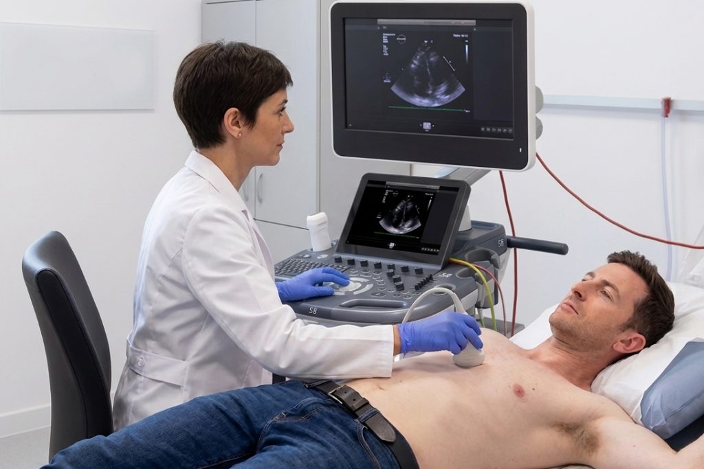
How is the test performed?
The examination is carried out in an outpatient medical consultation and does not require hospital admission. The patient lies on the examination table with the upper body uncovered. The cardiologist applies conductive gel to the chest area and moves the echocardiography probe across the chest surface to obtain the necessary images. During the examination, some position changes may be required, which the doctor will indicate. The test usually lasts between 15 and 30 minutes.
Is it painful or dangerous?
No. A cardiac ultrasound is a completely safe and painless test. It is based on ultrasound waves, so it does not use radiation or any external agents. It does not damage tissues and can even be performed in pregnant women. Most patients experience no discomfort during the examination.
Do I need special preparation or to fast beforehand?
No prior preparation is necessary. Patients can attend the appointment in their usual condition, without fasting, without stopping medication, and without any special restrictions before the test.
Do I need a doctor’s referral or can I have an echocardiogram privately?
Yes, you can request the test without a referral. At our clinic, the test includes an immediate cardiology assessment by the specialist, so presenting a prior medical order is not mandatory.
However, although you may book an appointment directly, it is advisable to provide a referral from your doctor if you have one; this helps us better understand your medical history and guide the diagnosis more accurately.
How is it different from an electrocardiogram?
These are different and complementary tests. An electrocardiogram (ECG) records the electrical activity of the heart and reflects its rhythm and conduction. An echocardiogram, on the other hand, provides a direct image of the heart in motion and allows visualization of its structure, wall thickness, valve function, and blood flow. In other words: the electrocardiogram shows how the heart functions electrically; the echocardiogram shows what the heart looks like and how it moves. In many cases, the cardiologist uses both tests together to obtain a complete assessment.
Is it recommended for athletes?
Yes, especially in cases where the sports medical examination or federation electrocardiogram shows abnormalities. An echocardiogram can rule out structural heart diseases that may pose a risk during high-intensity sports practice. It is a common test in sports cardiology evaluations and helps ensure safe physical activity.
What is its relationship with the Holter monitor?
The Holter monitor and the echocardiogram are different and complementary cardiology tests. A Holter monitor is a device that continuously records electrical activity or blood pressure over 24 hours, making it particularly useful for detecting intermittent arrhythmias. An echocardiogram, on the other hand, provides information about the structure and mechanical function of the heart. Depending on the clinical case, the cardiologist will determine whether one or both tests are necessary.